Abstract
Background: The classical BCR-ABL1-negative Myeloproliferative Neoplasms (MPNs) are clonal haemopoietic stem cell disorders, include Primary Myelofibrosis (PMF), Polycythemia Vera (PV) and Essential thrombocythemia (ET).The lack of specific discriminator makes the subcategorization of BCR-ABL1 negative Myeloproliferative Neoplasms very challenging, especially in distinguishing ET from prefibrotic MF cases. The emphasis on the need for an accurate diagnosis is because of differences in clinical outcome and therapy.
Recently, h-MICL (also termed CLEC12A, CLL-1, or CD371) is emerging as a promising marker of leukemic stem cells (LSC) in AML. Its most prominent features is its lack of expression on CD34+CD38-cells in normal bone marrow, regenerating bone marrow and G-CSF stimulated peripheral blood stem cells, making it a highly specific marker for LSCs. Recent studies claim that its specificity for LSCs makes it a promising target for future targeted therapy as well. Thus, h-MICL can turn out to be a crucial marker in the diagnostics of MPN for discriminating MPN phenotypes.
Aims & Objectives:
To evaluate the utility of flowcytometric enumeration of circulating CD34+CD38- hMICL+ cells in subcategorisation of BCR-ABL1 negative MPNs.
To define a discriminatory cut-off of aberrant h-MICL expression at the stem cell level for accurate identification of PMF patients and to especially differentiate early prefibrotic PMF and ET.
To correlate aberrant h-MICL+ stem cell subset with Dynamic International Prognostic scoring System (DIPSS score).
Materials & Methods:
A total of 54 BCR-ABL1 negative MPN patients, including 15 PV, 13 ET ,13 Overt MF, 8 early prefibrotic stages of Primary MF, and 5 Post-PV/ET MF, who presented in the clinics of Hematology department of a tertiary care center in North India were included in the study. Bone marrow examination and mutational analysis were carried out as part of diagnostic workup.
Peripheral blood flowcytometric evaluation was done. The samples were processed using stain-lyse-wash method. Flowcytometric analysis was done on BD FACS Canto II (Becton Dickinson, San Jose, CA, USA) using BD FACSDiva™ v8.1.3 software.The subsets analysed were:
CD34+ (hematopoietic stem cell enriched population)
CD34+CD38+ (committed progenitors)
CD34+CD38- (stem cells)
The aberrant CD34+CD38-hMICL+ population
The aberrant hMICL expression on CD34 + CD38- subsets were correlated with DIPSS score too.
Results:
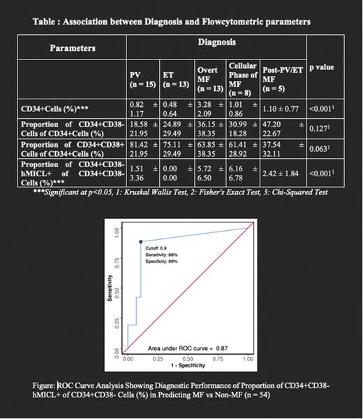
Our study included 54 BCR-ABL1 negative MPN patients (42 males, 12 females) with a median age of 49.5 years. A significant difference in distribution of proportion of CD34+CD38-hMICL+ among the MPN sub-groups was found. The median of CD34+CD38-hMICL+cells subset in Overt MF, early stage/ pre PMF and Post-PV/ET MF group was 2.8%, 4.15% and 3% respectively, which was significantly more than PV and ET (p<0.001).
Discriminatory cut off of Proportion of CD34+CD38-hMICL+ subset ≥0.9 predicted MF with sensitivity and specificity of 88% and 89%respectively. Cut off to differentiate early/ prefibrotic MF from ET was found to be ≥2 (sensitivity:87.5%;specificity:100%)
We found a statistically significant correlation (rho = 0.41, p = 0.036) between proportion of CD34+CD38-hMICL+ cells and DIPSS score. For every 1 unit increase in DIPSS Score, CD34+CD38-hMICL+ cells increased by 1.72%.
Discussion & Conclusion:
Subcategorization of BCR-ABL1 negative MPNs by morphology alone can be perplexing due to overlapping features and interobserver reproducibility. Driver mutations do not differentiate subtypes. In this study we conclude that CD34+ CD38- hMICL+ subset is reliable for subcategorization of BCR ABL1-negative MPNs. We defined a cut-off to discriminate MF from non-MF MPNs, also prefibrotic MF from ET. To the best of our knowledge, there is only one similar study by Herborg LL, et al (2018). They proposed a cut off of >0 cells as discriminator, with 80% sensitivity and 97% specificity. However, they did not distinguish early stage/prefibrotic PMF and Post-PV/ ET MF. Moreover, study of the correlation of aberrant hMICL expression with any prognostic scoring system is not done. Hence our study is unique and very first of its kind in this regard. We conclude that flowcytometric enumeration of circulating CD34+CD38- h-MICL+ cell subset holds a potential as a robust diagnostic test, tool for prognostication, an indicator of disease progression and a potential target for therapy.
Disclosures
Seth:Takeda: Honoraria.
Author notes
Asterisk with author names denotes non-ASH members.


This feature is available to Subscribers Only
Sign In or Create an Account Close Modal